Ultrasound Imaging in Los Angeles

Sometimes referred to as sonography or medical sonography, diagnostic ultrasound utilizes high frequency sound waves above the range of human hearing to produce images of internal structures.
There are many reasons to schedule an ultrasound. This remarkable technology, particularly 3d ultrasound, is now routinely used to chart the course of a pregnancy and to determine the health and sex of the baby. Ultrasound is one of the most common diagnostic tools used today, responsible for averting life-threatening conditions that were formerly hidden to physicians.
Los Angeles is home to a world-class medical community including some of the top hospitals in the country. These professionals rely on precise information from ultrasound and other diagnostic equipment to assist them in fighting disease and restoring health. One of the greatest benefits from having reliable and precise information is achieving peace of mind. Knowledge is crucial to successful treatment of any medical condition. Knowing there is nothing to worry about is priceless.
Latest technology - 3D & 4D ultrasound Los Angeles
 Optima Diagnostic Imaging takes pride in providing the latest diagnostic equipment in partnership with top medical technicians fully trained and up to date on the latest improvements in the equipment. In this way, they provide top tier 4d and 3d ultrasound in Los Angeles many thousands of times each year.
Ultrasound procedure
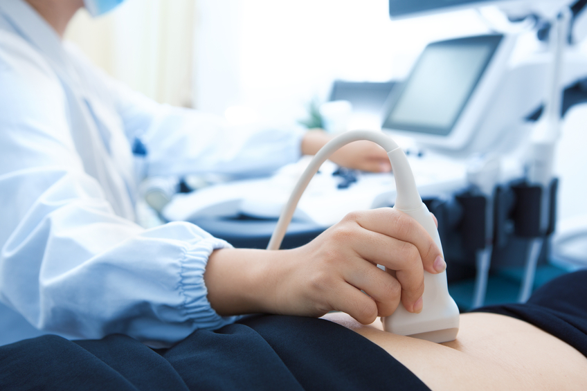
The technician administering an ultrasound uses a small handheld transducer that delivers sound waves into the body. The sound waves return to the device and are delivered into a computer, which interprets the waves into a viewable image. These images provide valuable data concerning internal organs and structures. Because an ultrasound utilizes sound waves instead of radiation, it is an entirely safe procedure.
There is virtually no preparation necessary to administer this type of imaging examination. There is no pain involved, only the administration of some gel to improve the clarity of the image.
How Ultrasound Imaging Works
In an ultrasound test, sound waves are sent into the targeted area of the body, and the echo of the sound waves is sent to a computer that creates a real-time image of the systems within the body structure. It is safe and painless, and does not involve the use of radiation.
Preparing for an Ultrasound Imaging Test
Prior to your ultrasound, you will be given directions about what you need to do to prepare. In many cases, all you will need to do is wear comfortable, loose-fitting clothing. Different areas of the body may require more specific pre-test instructions. An ultrasound performed on the abdomen may require you to eat a fat-free meal the evening prior to the test, to avoid food or drink for 12 hours prior to the test, and to drink a quart of water one hour prior to the test so your bladder is full, to help create a better image. Many health insurance plans cover all or part of the cost of ultrasound image tests.
The Ultrasound Test
For the test, you will be asked to remove some (or all) of your clothing, depending upon the area of the body to be viewed. Gel is spread on the skin of the area to be tested. The image is produced with the use of a hand-held device called a transducer, which the technician moves back and forth over the body. Images are displayed on a screen, and these images are saved so they can be reviewed by a radiologist to aid in diagnosis. In some cases, a probe is inserted in the body, such as in cases of suspected prostate cancer or to view the uterus or ovaries in a woman. The test will take only 20 minutes to an hour to perform.
Ultrasound at Los Angeles Optima Diagnostic Imaging include:
- Neck/Thyroid
- Chest
- Renal
- Testicular with Duplex
- Breast
- Abdomen
- Abdomen Doppler
- Aorta Duplex (AAA)
- Pelvic
- Carotid Duplex
- Venous Duplex (Lower and Upper Extremities)
- Arterial Duplex (Lower and Upper Extremities)
- Ankle Brachial Index (ABI)
- Hemodialysis Graft Evaluation
- Vein Mapping
We offer 4D and 3D ultrasound imaging in Los Angeles. Read below to find out more about each type of ultrasound imaging we provide.
Neck/Thyroid Ultrasound
The thyroid is the gland in the neck that regulates metabolism. A thyroid ultrasound creates images of this area to locate any benign or malignant thyroid tumors.
Renal Exam Ultrasound
This is an ultrasound of the kidney. It can detect any masses in the kidney, such as kidney stones or cysts.
Testicular with Duplex Ultrasound
This is an ultrasound of your testicles. Like all our ultrasound exams, it is a painless procedure, allowing doctors to see different types of tissues and diagnose a variety of medical conditions.
Breast Ultrasound
This produces an image of the inside of the breast. It can be used to determine if any abnormality is solid or fluid-filled, such as in non-cancerous lumps or cysts.
Abdomen Ultrasound with Doppler
This procedure uses the Doppler, which allows the ultrasound specialists to evaluate blood flow through the arteries and veins in your abdomen. This allows the ultrasound specialists to see any obstructions in the blood flow to your abdominal organs and diagnose what the obstruction is. It can also detect conditions including gallbladder disease, gallstones, liver, pancreas, kidney and spleen problems and masses in your abdomen.
Abdomen Ultrasound
This procedure allows the ultrasound specialists to diagnose medical conditions such as gallbladder disease, gallstones, liver, pancreas, kidney and spleen problems and masses in your abdomen.
Aorta Duplex
This procedure is used to evaluate the aorta, the main vein that runs through your body, to detect any aneurysms. This is commonly done for people with diabetes, high blood pressure and high cholesterol. Those issues can increase the risk of someone getting an abdominal aorta aneurysm.
Pelvic Ultrasound
This ultrasound can help determine what is causing pain or bleeding in reproductive organs.
OB Ultrasound
The OB ultrasound is an obstetric ultrasound, which produces an image of a fetus inside a woman’s uterus, as well as the mother’s ovaries and uterus. An OB ultrasound is commonly used to evaluate various factors of the fetus’s health, including growth and positioning. It also can help determine things such as the amount of amniotic fluid around the baby, the position of the placenta and the overall well being of the baby.
The OB ultrasound is done at different times during the pregnancy to continue to monitor the health throughout the pregnancy and to diagnose any issues that may arise. This is commonly done as a 3D ultrasound for patients who want to get a three-dimensional look at their baby.
Fetal Doppler Ultrasound
This is used to detect the heartbeat of a fetus and determine any prenatal care that may be needed.
Nuchal Translucency Ultrasound
The nuchal translucency ultrasound is used to screen for Down syndrome and other congenital heart defects in a fetus.
Carotid Duplex Ultrasound
The carotid duplex ultrasound is used to examine the how the carotid arteries in your neck are functioning. The carotid arteries are on either side of your neck and are the veins that bring blood from your heart to your brain. This ultrasound can help detect narrowed carotid arteries, which can increase the risk of a stroke.
Ultrasound Options In Los Angeles
To learn more about our many ultrasound options, call our office today. We provide many options for ultrasounds in Los Angeles, including 3D & 4D ultrasounds that can show you the face of your baby or track blood flow through your arteries. Ultrasound tests are a great, safe method of monitoring and diagnosing health conditions and are truly a remarkable window into the internal mechanics of the body. Our technicians here in Los Angeles are trained and ready to help you.
Ultrasound Testing at Beverly Hills Cancer Center
Ultrasound imaging allows doctors to identify a tumor, as well locate the exact area upon which to perform a biopsy. At Beverly Hills Cancer Center, we use the most advanced testing methods and technology in our state-of-the-art facility, including ultrasound imaging.
Identifying tumors with ultrasound imaging
The ultrasound imaging test is used to identify tumor growth in various areas of the body. Once an area is identified, a biopsy can be performed to test the tissue for cancer cells. This is termed “image-guided” biopsy. These tests are frequently ordered to identify cancer growths in the lymph nodes, breasts, and liver or other parts of the body. Ultrasound can also be used in combination with endoscopy (a small flexible tube with a tiny camera inserted in the body to view certain areas) or bronchoscopy (a flexible tube inserted through then nose and down the throat with a camera for viewing the lungs) to identify tumors in the gastrointestinal tract or lungs.
Ultrasound FAQ:
What is the Difference Between 3D & 4D Ultrasounds?
A 3D ultrasound will produce a three-dimensional image that provides a more comprehensive, detailed image than a simple 2D ultrasound. Meanwhile, a 4D ultrasound looks three-dimensional but will include movement.
Are There Any Risks with Ultrasound?
As opposed to other kinds of imaging, ultrasound tests do not emit radiation. The same technology is used for all types of ultrasound tests with no adverse effects discovered, making it a safe and painless procedure that is useful for virtually everyone.
Can I Still Take Medications Before my Ultrasound?
Yes, you can still take medications before your ultrasound. Some types of ultrasounds will require a short fast, and any medications should only be taken with a small amount of water during that time. Otherwise, there will be little to no change to your diet or medications.
When Will I Get my Ultrasound Results Back?
Usually your results will be available right away. Sometimes the findings are obvious and will be discussed at once, and in some situations the doctor will need more time to analyze the images and come up an accurate diagnosis.
How Long Will my Ultrasound Take?
Your ultrasound will likely take about 30 minutes, although the examination can vary, based on the area being analyzed and the amount of detail needed.
Are There Limitations to Ultrasound Imaging?
Ultrasound waves are easily disrupted, so an ultrasound is not ideal for looking at air-filled organs or patients who are overweight. Ultrasound tests are best used for soft tissues, not internal structure of bones.
What Impacts the Image Quality of an Ultrasound?
The more tissue the ultrasound waves need to travel through, the more they can be distorted. Your ultrasound image may be negatively impacted by:
- The presence of air in your organs
- Layers of fat cells
- The positioning of your organs
